Significance
Biological reactions are strongly coupled to the structure and properties of their environment. Cellular compartmentalization into biomolecular condensates with distinct material properties enables a diverse array of biological processes. However, the link between material properties and function is challenging to study due to the difficulties of measuring intracellular condensate material properties. Here, we probe the material properties of a biochemically active condensate, the nucleolus, inside a living cell nucleus using micropipette aspiration (MPA). We find that the nucleolus is more solid-like at its core and more liquid-like toward its periphery. These properties are RNA-dependent; by degrading RNA, the nucleolar core becomes more liquid-like. These results provide insight into how nucleolar material properties may be linked to the sequential steps of ribosome maturation.
Abstract
The nucleolus is a multiphasic biomolecular condensate that facilitates ribosome biogenesis, a complex process involving hundreds of proteins and RNAs. The proper execution of ribosome biogenesis likely depends on the material properties of the nucleolus. However, these material properties remain poorly understood due to the challenges of in vivo measurements. Here, we use micropipette aspiration (MPA) to directly characterize the viscoelasticity and interfacial tensions of nucleoli within transcriptionally active Xenopus laevis oocytes. We examine the major nucleolar subphases, the outer granular component (GC) and the inner dense fibrillar component (DFC), which itself contains a third small phase known as the fibrillar center (FC). We show that the behavior of the GC is more liquid-like, while the behavior of the DFC/FC is consistent with that of a partially viscoelastic solid. To determine the role of ribosomal RNA in nucleolar material properties, we degrade RNA using RNase A, which causes the DFC/FC to become more fluid-like and alters interfacial tension. Together, our findings suggest that RNA underlies the partially solid-like properties of the DFC/FC and provide insights into how material properties of nucleoli in a near-native environment are related to their RNA-dependent function.
The interior of living cells is compartmentalized into both membrane-bound and membrane-less organelles. Many membrane-less organelles, from nucleoli and Cajal bodies in the nucleus to P-bodies and stress granules in the cytoplasm, form through phase separation and related phase transitions, with the resulting condensed assemblies of proteins and RNAs referred to as biomolecular condensates . The kinetic and thermodynamic driving forces underlying condensate formation can give rise to a wide range of material states, from liquid-like to gel or solid-like. These condensate material states are correlated with a variety of biological functions , and aberrant changes in condensate material state are associated with biological dysfunction and disease. For example, condensate liquid-to-solid transitions are linked to various neurodegenerative diseases, including those driven by FUS , tau , polyQ , alpha-synuclein , hnRNPA1 , and other proteins.
Condensate material states can be characterized in terms of macroscopic material properties, which include viscosity, elasticity, and interfacial tension . A major challenge in the condensate field is the difficulty of in vivo measurements of these material properties. While many rheological techniques are capable of measuring such properties with in vitro condensates, these reconstituted systems are vast simplifications of native condensates in their cellular context. Existing methods for inferring condensate material properties in vivo rely on indirect measurements and often use condensates containing overexpressed, fluorescently tagged proteins, which can give rise to altered material properties. Within living cells, condensate viscosity has been very roughly estimated from measurements of the diffusion coefficient of fluorescently labeled condensate proteins using fluorescence recovery after photobleaching (FRAP) or fluorescence correlation spectroscopy (FCS) (. However, the diffusion of one protein species may not fully reflect the bulk rheology of the whole condensate, which could contain hundreds of different protein species, and assumptions of molecular size, shape, and material homogeneity are needed to convert the measured diffusion coefficient to condensate viscosity.
Another material property of interest is interfacial tension (, which in principle can be estimated from the fluctuations in droplet shape (, but for condensates also typically requires fluorescently labeled proteins, and is challenging due to the small scale of typical fluctuations . The relaxation time of coalescing condensates has also been used to approximate the ratio between viscosity and interfacial tension (inverse capillary velocity) , but must be paired with another technique to extract viscosity, which can usually only be estimated roughly. An additional challenge is that these techniques require assuming that condensates are liquid-like, Newtonian fluids , even while many condensates appear to exhibit characteristics of gel- or solid-like materials. For instance, condensates with proteins that exhibit incomplete FRAP recovery , or those that do not dissolve following salt/chemical treatment , are thought to be more solid-like, although these techniques only provide qualitative observations.
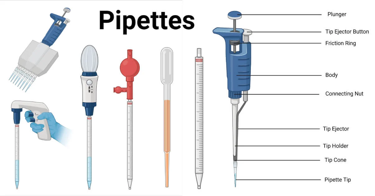
Given the limitations of current techniques used to characterize the viscoelastic and surface properties of condensates in cells, there is a need for approaches that can directly measure in vivo condensate material properties. Micropipette aspiration (MPA) is a well-established, label-free technique for probing the mechanical properties of cells , and has recently been applied to study the viscoelasticity and interfacial tension of in vitro condensates . MPA measures a one-dimensional strain in response to an applied stress, which takes the form of the pressure drop imposed across the pipette. The minimum pressure required for a condensate to enter the pipette is a direct measure of interfacial tensio. Once inside the pipette, generally, a fluid-like, Newtonian material will deform with a constant strain rate ε˙ under applied constant stress, σ, i.e., σ=ηε˙, while a solid-like material deforms with a constant strain, ε, i.e., σ=Eε, where η and E are, respectively, the viscosity and elastic modulus of the material. Viscoelastic materials display strain responses consistent with both fluid-like and solid-like behaviors at short times, but may generally be classified into fluid-like and solid-like based on their long-time behavior. Because MPA requires condensates larger than the micropipette opening diameter, adapting MPA for studying intracellular condensates, especially those inside the cell nucleus, is challenging due to the small size of typical condensates .
To leverage the full power of MPA for measuring condensates in cells, we study the large nuclear condensates within Xenopus laevis oocytes, which are structurally and compositionally similar to those in typical mammalian systems . The potential for deploying MPA and related techniques for measuring native condensate properties in X. laevis oocytes is particularly attractive for the nucleolus — the largest and still elusive multiphasic biomolecular condensate responsible for the production of ribosomes, highly complex assemblies composed of ~80 proteins and four ribosomal RNAs (rRNAs) in eukaryotes . Three rRNAs are transcribed within the nucleolus, where they are modified, and assembled with dozens of proteins into precursor ribosomal subunits. These processing steps are spatially organized within the nucleolus in three concentrically arranged subcompartments, with the fibrillar center (FC) as the innermost compartment, followed by the dense fibrillar component (DFC) and granular component (GC). The nucleolus exhibits liquid-like material behaviors (, and the proper execution of ribosome biogenesis may be coupled to these properties. For example, inducing nucleolar gelation through oligomerization of the GC protein, nucleophosmin (NPM1), leads to the accumulation and depletion of different precursor rRNAs. Moreover, the transcription and processing of rRNA may modulate the material properties of the individual phases of the nucleolus. Indeed, newly transcribed rRNAs in the DFC have been proposed to entangle and form a gel-like state, while rRNAs that have been packaged and assembled into preribosomal subunits in the GC could contribute to a more liquid-like state . The potential for significant RNA-dependent viscoelasticity in the nucleolus would be consistent with previous measurements on the incomplete FRAP recovery of FIB1-eGFP . However, the lack of direct measurements of nucleolar material properties severely limits our understanding of these properties and the potential contributions of rRNAs.
Here, we use MPA to directly characterize the viscoelastic properties and interfacial tensions of nucleoli within transcriptionally active X. laevis oocytes and elucidate the role of RNA in influencing these material properties. We show that the major nucleolar compartments, the GC and DFC/FC, have distinct material responses. Specifically, the GC is liquid-like, while the DFC/FC is more solid-like. We further show that these differences arise largely from RNA, whose degradation fluidizes the DFC.
Results
Fluid Behaviors of Nucleoli During MPA.
To probe the material properties of the nucleolus, we take advantage of the large ~10 μm nucleoli of stage V-VI X. laevis oocytes, which are single cells that reach diameters of 1 to 1.3 mm. The nucleus of each oocyte, known as the germinal vesicle (GV), contains 1,400 to 1,600 nucleoli that form around extrachromosomal rDNA repeats and are suspended within an actin meshwork . X. laevis nucleoli share a similar tripartite organization with mammalian cells, containing nested FC, DFC, and GC compartments. While nucleoli can be visualized without fluorescent labeling by differential interference contrast (DIC) microscopy, to distinguish the different nucleolar subcompartments, we label the GC and DFC by expressing fluorescently tagged nucleophosmin (NPM1-RFP) and fibrillarin (FIB1-eGFP), respectively. Because the oocyte cytoplasm is opaque, we isolate the GVs into mineral oil to facilitate imaging . As previously described, these nucleoli exhibit liquid-like behaviors and disrupting the actin meshwork causes sedimentation under gravity and apparently liquid-like fusion of the GC and DFC


